- Mon - Sat 9:00am - 9:00pm
- Sun 9:00am - 2:00pm
- No 218, South Service Road, Phase1,
- Sathuvachari, Vallalar, Vellore - 632009
- Landmark: Near Vallalar Bus Stop
Head & Neck

Cleft Lip
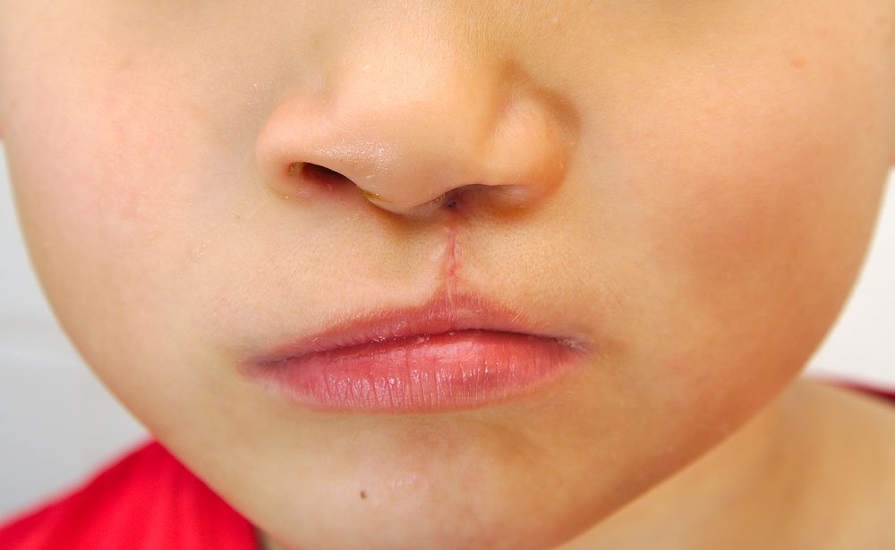
A unilateral cleft lip may appear as a small notch in the edge of the lip only or extend into the nose or gums. A child may be born with just a cleft lip or may have a cleft palate as well, which is a split in the roof of the mouth. Sometimes a cleft occurs as part of a syndrome, meaning there are other birth defects. Other times, it's genetic and runs in families. A cleft also can be associated with environmental factors, such as a woman's use of certain medications, exposure to cigarette smoke, or lack of certain vitamins while pregnant. Most of the time, though, the cause isn't known. Cleft lip can be associated with other problems, including feeding difficulties, fluid buildup in the middle ear and hearing loss, dental abnormalities, and speech difficulties.

Cleft palate
An opening in the roof of the mouth due to a failure of the palatal shelves to come fully together from either side of the mouth and fuse during the first months of development as an embryo. The opening in the palate permits communication between the nasal passages and the mouth. Surgery is needed to close the palate. Cleft palate can occur alone or in association with cleft lip. During the first 6 to 10 weeks of pregnancy, the bones and skin of a baby's upper jaw, nose, and mouth normally come together (fuse) to form the roof of the mouth and the upper lip. A cleft palate happens when parts of the roof of the mouth do not fuse together completely.
Branchial Sinus/Fistula
During early prenatal development, gill-like structures (branchial) usually resorb but in rare circumstances, they may remain. These structures may connect with the skin only and drain sloughed skin through a small opening on the skin (branchial sinus); with the skin and the throat lining and drain mucous through a small opening on the skin (branchial fistula); or have no connection at all and slowly grow over time (branchial cyst). All of these are referred to as branchial anomalies. Branchial anomalies typically are present on the front of the neck, but may be seen anywhere from the lowest portion of the neck, the thyroid gland or to the area around the ear. Branchial anomalies may be present at birth, or may become enlarged during or following an upper respiratory tract infection.
Neck Cysts
Neck cysts are a common problem for infants and children, are usually benign masses, and may be present at birth. Common types are: Branchial cleft abnormalities: These tissues may form cysts (pockets that contain fluid) or fistulas (passages that drain to an opening in the skin surface). They are usually located near the front edge of the neck muscle that goes from the jawbone across to the collarbone and breastbone. Thyroglossal duct cysts: These neck masses or lumps develop from cells and tissues remaining after the formation of the thyroid gland during embryonic development. This type of cyst tends to be diagnosed in preschool-aged children or in mid-adolescence and often appears after an upper respiratory infection, when it enlarges and becomes painful.

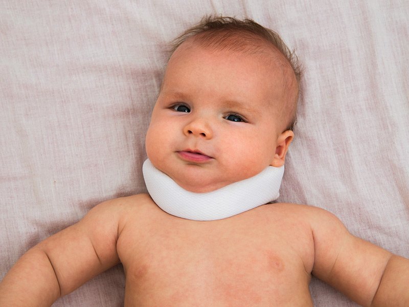
Torticollis
Torticollis is a problem involving the muscles of the neck that causes the head to tilt down. The term comes from two Latin words: tortus, which means twisted, and collum, which means neck. Sometimes it’s called “wryneck.” If your baby has the condition at birth, it’s called congenital muscular torticollis. That’s the most common type. Babies can also develop the condition after birth. Then it’s called “acquired,” rather than congenital. Acquired torticollis may be linked to other, more serious medical issues.